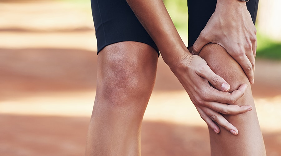
What are common signs of Patellar Tendonitis?
- Pain and tenderness in the front of the knee, especially where the tendon attaches to the kneecap
- Stiffness in the knee, especially after sitting for a long period of time
- Swelling or redness in the knee
- A crackling or popping sensation in the knee when moving
- Decreased range of motion in the knee
- Weakness in the knee, making it difficult to perform activities that put stress on the knee, such as jumping and running
If you experience any of these symptoms, it’s important to see a doctor for a proper diagnosis and treatment plan. Left untreated, patellar tendonitis can become a chronic condition and lead to more serious problems.
How do you get patellar tendonitis?
Patellar tendonitis, also known as jumper’s knee, is caused by repetitive stress and strain on the patellar tendon, which connects the kneecap (patella) to the shin bone (tibia). Activities that put a lot of strain on the knee, such as jumping, running, and playing sports, can lead to patellar tendonitis. Other factors that may increase the risk of developing the condition include:
- Tight or weak muscles in the legs
- Poor training techniques or overtraining
- Improper footwear
- Inflammation from arthritis
It’s important to treat patellar tendonitis early to avoid further damage to the tendon and to reduce pain and discomfort.
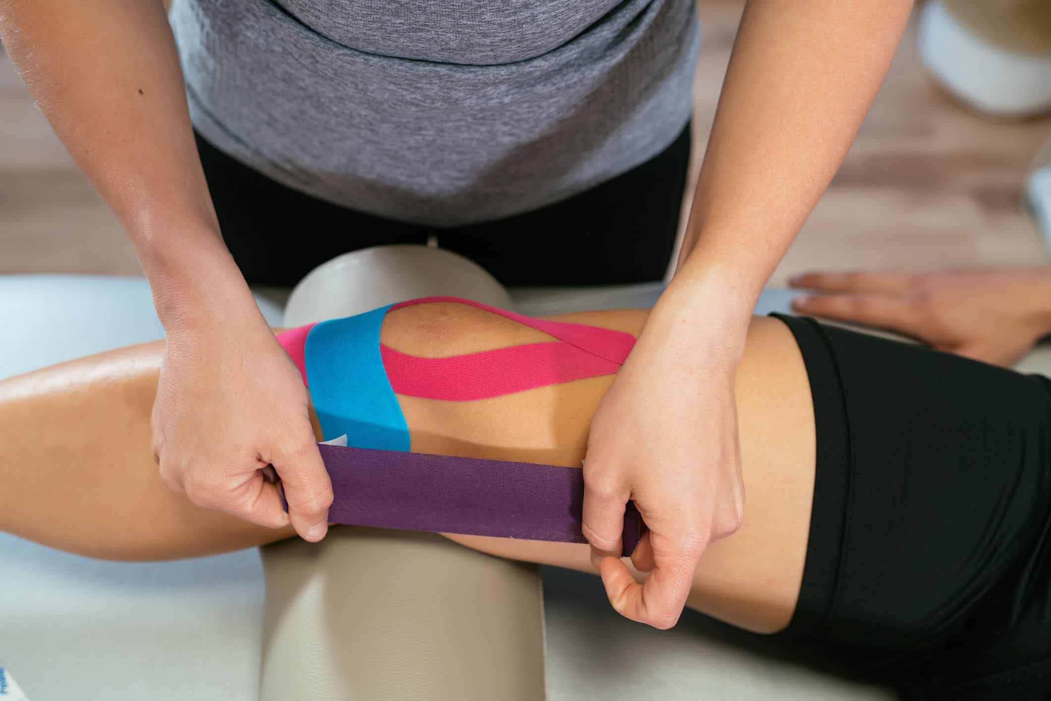
What can you do for patellar tendinitis?
There are several treatment options for patellar tendinitis, including:
- Rest: Avoiding activities that cause pain and discomfort can help reduce inflammation and allow the tendon to heal.
- Ice: Applying ice to the affected area can help reduce pain and swelling.
- Physical therapy: Strengthening and stretching exercises, as well as other physical therapy techniques, can help improve the flexibility and strength of the knee, reducing the risk of reinjury.
- Nonsteroidal anti-inflammatory drugs (NSAIDs): Over-the-counter pain relievers such as ibuprofen can help reduce pain and inflammation.
- Orthotics: Custom-made shoe inserts can help distribute weight evenly and reduce stress on the knee.
- Steroid injections: In some cases, a doctor may recommend a corticosteroid injection to reduce inflammation and pain.
- Surgery: In severe cases, surgery may be necessary to repair the damaged tendon.
It’s important to consult a doctor to determine the best treatment plan for your individual case.
What is your patellar tendon?
The patellar tendon is a strong, fibrous tissue that connects the patella (knee cap) to the tibia (shin bone) in the lower leg. It plays an important role in transmitting force from the quadriceps muscle to the tibia, allowing for movement and stability in the knee joint. The patellar tendon helps to straighten the leg, and also acts as a shock absorber during physical activities such as jumping and running. Pain and inflammation in the patellar tendon is a common condition known as patellar tendinitis or “jumper’s knee”.
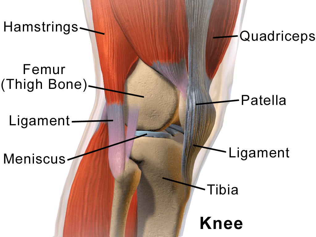
Anatomy of your knee:
The knee is a hinge joint located at the intersection of the thigh bone (femur) and the shin bone (tibia). It is the largest joint in the body and allows us to bend and straighten the leg. The following are some of the main anatomical structures of the knee:
- Tibia: Also known as the shin bone, the tibia is one of the two bones that make up the lower leg. It provides stability to the knee joint and supports the weight of the body.
- Femur: The femur, or thigh bone, is the longest and strongest bone in the body. It connects the hip to the knee and provides the main source of power for leg movement.
- Patella: Also known as the kneecap, the patella is a small, triangular bone that covers and protects the knee joint. It is connected to the quadriceps muscle by the patellar tendon.
- Menisci: The menisci are two crescent-shaped pieces of cartilage that sit between the femur and tibia. They help distribute weight evenly across the knee and provide shock absorption.
- Ligaments: The knee has four main ligaments that provide stability and support to the joint: the anterior cruciate ligament (ACL), the posterior cruciate ligament (PCL), the medial collateral ligament (MCL), and the lateral collateral ligament (LCL).
- Bursae: Bursae are small, fluid-filled sacs that help reduce friction between bones, tendons, and muscles in the knee.
- Synovial membrane: The synovial membrane is a thin layer of tissue that surrounds the knee joint and produces synovial fluid, which helps lubricate the joint and reduce friction.
Looking for Physiotherapy in Langley? Be sure to checkout Revamp Wellness for all of your rehabilitation needs!
https://rvwlangley.janeapp.com/
references:
OpenAI. (2021). OpenAI. Retrieved from https://openai.com/